Mammography: When You Should Have a Breast Examination and What It Can Detect
Reviewed by Victor Vladimirovich Khanov, Oncosurgeon of the highest category. Mammologist. General Director updated: 16 July 2025
Breast health requires regular attention throughout a woman's life. Many breast diseases develop without noticeable symptoms in their early stages, making timely diagnosis one of the most effective ways to detect them. Mammography is considered the gold standard for breast imaging because it can identify even the smallest abnormalities before clinical symptoms appear. Regular screening allows treatment to begin at an early stage and significantly improves the chances of a full recovery.
What Is Mammography and Why Is It Performed?
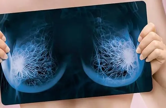
Mammography is an X-ray examination of the breasts used for the early detection of both benign and malignant breast conditions. During the procedure, high-resolution images of the breast tissue are obtained, allowing the physician to evaluate its structure and identify lumps, calcifications, cysts, and other abnormalities.
Mammography is widely used both as a preventive screening tool and as a diagnostic examination for patients with symptoms or increased risk factors. Thanks to modern imaging equipment, radiation exposure is minimal while diagnostic accuracy remains very high.
If abnormalities requiring surgery are detected, modern breast surgery techniques may be recommended. Learn more about breast surgery follow the link
When Should You Have a Breast Examination?
Routine screening mammography is recommended for all women over the age of 40, even in the absence of symptoms. The frequency of examinations is determined by the physician based on age, family history, overall health, and individual risk factors.
You should schedule a medical consultation if you notice:
- a lump in the breast
- breast pain or discomfort
- changes in breast size or shape
- nipple retraction
- nipple discharge
- redness or swelling of the breast skin
- enlarged lymph nodes in the armpit.
Mammography may also be recommended for women with a family history of breast cancer or for follow-up after treatment of breast diseases.
How Is Mammography Performed?
Mammography requires little preparation. The examination is usually scheduled during the first half of the menstrual cycle, when breast tissue is generally less sensitive.
During the procedure, each breast is placed between two plates of the mammography unit, and several X-ray images are taken from different angles. Gentle compression of the breast tissue is necessary to produce the clearest possible images while minimizing radiation exposure.
The examination typically takes 10–15 minutes. Most women experience only mild temporary discomfort, which disappears shortly after the procedure. Once the images have been analyzed, the physician prepares a report and, if necessary, recommends additional diagnostic tests.
What Can Mammography Detect?
Mammography helps identify a wide range of breast conditions before symptoms become apparent. It can detect:
- benign breast tumors
- cysts
- fibroadenomas
- intraductal abnormalities
- calcifications
- fibrocystic breast disease
- inflammatory conditions
- precancerous lesions
- malignant breast tumors.
Mammography plays a particularly important role in the early detection of breast cancer. Early diagnosis significantly improves treatment success and patient outcomes. If breast cancer is confirmed, further treatment is planned by a multidisciplinary team using modern oncologic surgery techniques. Learn more about oncologic surgery follow the link
Prevention and the Importance of Regular Screening
Regular mammography is one of the most effective methods for preventing serious breast diseases. In addition to routine screening, women are encouraged to perform monthly breast self-examinations, monitor for any changes, and seek medical advice promptly if any concerning symptoms develop.
Maintaining a healthy lifestyle, controlling body weight, staying physically active, avoiding smoking, and limiting alcohol consumption also contribute to better overall health. However, no preventive measure can replace regular medical screening.
Timely mammography helps detect breast diseases at an early stage, when treatment is most effective. Regular screening examinations and careful attention to your health can reduce the risk of complications and help maintain a high quality of life.

Was this article helpful?
Thank you for rating.
Prices
Schedule an appointment with a doctor


Frequently asked questions
History taking:
Studying the history of diseases and risk factors.
Examination and palpation:
Checking the mammary glands for changes, lumps and other abnormalities.
Diagnosis:
Ordering additional examinations (ultrasound, mammography, biopsy) if necessary.
Recommendations:
Determining treatment and further steps.
Still have questions?
Leave your details and a doctor will call you back.