
Lung CT Scan: When It Is Recommended and What the Examination Can Reveal
Computed tomography (CT) of the lungs is one of the most accurate diagnostic methods used in modern medicine to evaluate respiratory diseases. Thanks to its high-resolution imaging capabilities, CT provides a detailed assessment of the lung tissue, bronchi, pleura, and mediastinum, helping detect pathological changes at an early stage and determine the most appropriate treatment strategy. Unlike conventional chest X-rays, CT produces cross-sectional images of the organs, significantly improving diagnostic accuracy.
At the Helyos Medical Center, CT examinations are performed using advanced imaging equipment, while the results are interpreted by experienced radiologists, ensuring accurate and reliable diagnostics.
What Is a Lung CT Scan?
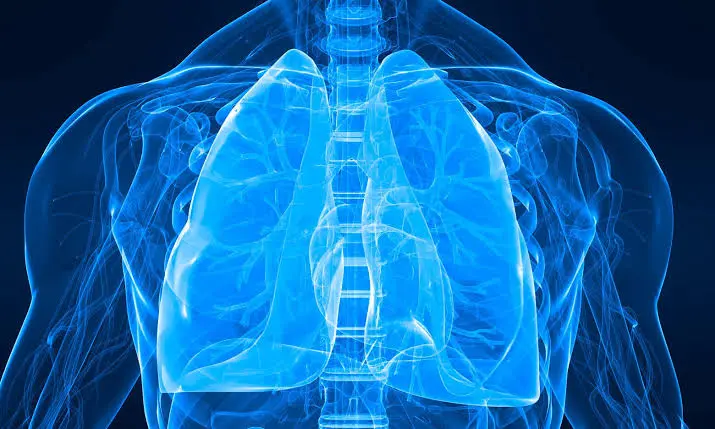
A lung CT scan is a non-invasive imaging technique that uses X-rays and advanced computer processing to produce detailed cross-sectional images of the organs within the chest.
During the examination, the scanner captures a series of rapid images from multiple angles. Specialized software then reconstructs these images into a detailed three-dimensional representation of the lungs and surrounding anatomical structures.
This technique allows physicians to detect even subtle abnormalities that may remain invisible on a standard chest X-ray.
When Is a Lung CT Scan Recommended?
A physician may recommend a CT scan of the lungs when respiratory disease is suspected or when additional information is needed to clarify the results of other diagnostic tests.
The main indications include:
- persistent cough;
- shortness of breath;
- coughing up blood (hemoptysis);
- chest pain;
- suspected pneumonia;
- prolonged fever;
- suspected tumors;
- follow-up after previous respiratory illnesses;
- evaluation of chest injuries.
Lung CT scans are also widely used before surgical procedures and to monitor the effectiveness of ongoing treatment.
What Conditions Can a Lung CT Scan Detect?
Computed tomography is one of the most informative imaging methods for evaluating the lungs.
A CT scan can help detect:
- pneumonia;
- chronic obstructive pulmonary disease (COPD);
- pulmonary emphysema;
- bronchiectasis;
- pulmonary fibrosis;
- tuberculosis;
- benign and malignant tumors;
- metastatic lesions;
- pleurisy (pleural inflammation);
- pulmonary embolism (when clinically indicated);
- congenital abnormalities of the lungs.
In addition, CT enables physicians to assess the condition of the lymph nodes, blood vessels, and mediastinal organs, making it an essential tool for the comprehensive evaluation of chest diseases.
How Is a Lung CT Scan Performed?
The examination is simple, non-invasive, and usually takes only a few minutes.
The patient lies on a movable table that slowly passes through the circular opening of the CT scanner. During the scan, it is important to remain still and briefly hold your breath when instructed by the medical professional.
In some cases, the examination is performed with an intravenous contrast agent, which provides better visualization of blood vessels and certain pathological changes.
Once the scan is completed, patients can usually return to their normal daily activities immediately unless their physician provides other instructions.
Preparing for a Lung CT Scan and Possible Contraindications
If the CT scan is performed without contrast enhancement, no special preparation is usually required.
Before a contrast-enhanced examination, your physician may recommend that you:
- avoid eating for several hours before the scan;
- undergo a blood test to assess kidney function;
- inform the medical team about any allergies;
- provide information about chronic medical conditions and current medications.
Possible contraindications include:
- pregnancy;
- severe allergic reactions to contrast agents;
- severe kidney impairment (for contrast-enhanced CT scans);
- certain serious medical conditions that make the procedure unsafe or impractical.
The decision to perform a CT scan is always made individually by the physician based on the patient's health status and clinical indications.
How Are CT Scan Results Interpreted?
After the examination, a radiologist carefully reviews the images and prepares a detailed diagnostic report.
The report typically includes an assessment of:
- the condition of the lung tissue;
- the presence or absence of abnormal lesions;
- changes in the bronchi;
- the condition of the pleura;
- the lymph nodes;
- the blood vessels and mediastinal organs.
Based on the CT findings, the treating physician may recommend consultation with a pulmonologist, additional diagnostic tests, or an appropriate treatment plan.
Conclusion
Computed tomography (CT) of the lungs is one of the most accurate diagnostic tools available for evaluating respiratory diseases. It enables the early detection of inflammatory conditions, tumors, vascular abnormalities, and many other clinically significant changes that are essential for selecting the most effective treatment strategy.
Timely CT examination, consultation with a qualified healthcare professional, and a comprehensive diagnostic approach can help initiate treatment at an early stage, improve clinical outcomes, and preserve long-term respiratory health.

Was this article helpful?
Thank you for rating.
Prices
Frequently asked questions
We recommend undergoing a CT scan if you have:
- prolonged or frequent headaches, dizziness, visual disturbances
- head, spine, or limb injuries
- suspected pneumonia, complications after COVID, or tuberculosis
- chest pain, shortness of breath, or coughing for no apparent reason
- abdominal pain of unknown origin
- suspected neoplasms
- follow-up after surgery or treatment
- desire to undergo a preventive examination
Types of computed tomography we perform
- CT scan of the brain
- CT scan of the chest
- CT scan of the abdomen and pelvis
- CT scan of the kidneys and urinary system
- CT scan of the spine (cervical, thoracic, lumbar)
- CT scan of the joints
- CT scan of the whole body
We offer conventional computed tomography and CT scans with contrast agent administration
How CT scans are performed at Helios:
Make an appointment by calling
Appointments at a convenient time without queues
Examination — you simply lie down while the tomograph works quietly and quickly
A radiology specialist interprets the results
Why choose Helios
State-of-the-art equipment — maximum image accuracy
Minimal radiation exposure
Fast results — in just a few minutes
Professional team of radiologists
No waiting in line
Comfortable atmosphere and friendly staff
At Helios, we care about diagnostic accuracy Your peace of mind and safety
Still have questions?
Leave your details and a doctor will call you back.